Echocardiography vs. ECG: Decoding the Rhythms and Structures of the Human Heart
Demystifying two common cardiac tests and understanding what each tells us about your heart.
When a patient presents with symptoms like chest pain, shortness of breath, or a racing heartbeat, assessing their cardiovascular health becomes an immediate priority. In these situations, physicians frequently order either an Electrocardiogram (ECG) or an Echocardiogram—and very often, they will order both. While the names sound similar, they are fundamentally different tests that serve entirely distinct diagnostic purposes.
Many patients confuse the two procedures, assuming that if they have had one, they do not need the other. To understand why both are often necessary, it is helpful to think of the heart as a house. An ECG is the test that checks the house's electrical wiring, while an Echocardiogram is the test that inspects the house's plumbing, walls, and doors.
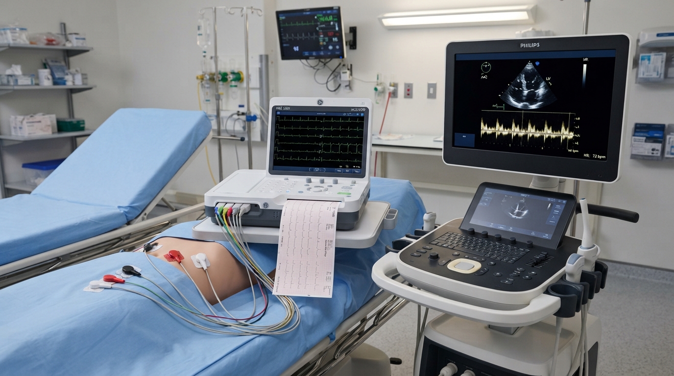
An Electrocardiogram (ECG or EKG) is a quick, painless test that measures the electrical activity of the heart. Every time the heart beats, an electrical signal travels through it, causing the muscle to contract and pump blood. Electrodes placed on the skin capture these signals and trace them on paper or a screen.
The ECG is the frontline tool for detecting arrhythmias (irregular heartbeats, whether too fast, too slow, or erratic). It is also the primary test used in emergency rooms to immediately identify if a patient is actively having a heart attack, or if they have had one in the past, by looking for specific changes in the electrical waveform.
However, an ECG tells us very little about the physical structure of the heart. It cannot tell us if the heart muscle is weak, if the valves are leaking, or if the heart is enlarged. For that structural information, we turn to the Echocardiogram.
An Echocardiogram is a specialized ultrasound of the heart. Using high-frequency sound waves, it creates real-time, moving images of the heart's chambers, walls, and valves. It allows the radiologist or cardiologist to literally watch the heart beating and pumping blood in real-time.
This visual data is incredibly rich. An echocardiogram can measure the 'ejection fraction'—the percentage of blood pumped out of the heart with each beat—which is a crucial indicator of heart failure. It can identify structural abnormalities, such as a leaky or stiff valve, congenital heart defects, or dangerous fluid accumulation around the heart (pericardial effusion).
Both tests are absolutely critical in the field of cardiology, and they complement each other perfectly. An abnormal electrical rhythm seen on an ECG might prompt a doctor to order an echocardiogram to see if a structural issue is causing the electrical problem.
Depending on your specific symptoms and medical history, your doctor will determine which test, or combination of tests, is necessary. At Crescent Radiology, our comprehensive cardiac imaging suite is fully equipped to perform detailed echocardiography, providing the accurate structural assessments your physician needs to keep your heart healthy.
Related Articles
Beyond Cholesterol: The New Era of Comprehensive Cardiovascular Risk Assessment
Read Article
Unmasking the Silent Killer: A Comprehensive Guide to Tracking and Taming Hypertension
Read Article