Empowering Women Through Early Detection: The Lifesaving Impact of Modern Mammography
Understanding the vital role of mammography and breast ultrasounds in the early detection of breast cancer.
Breast cancer remains the most commonly diagnosed cancer among women globally, casting a long shadow of anxiety and fear. However, the narrative surrounding breast cancer is rapidly changing from one of despair to one of hope and survival. The key driver behind this positive shift is early detection, primarily achieved through routine medical screening.
When breast cancer is detected in its earliest stages—before it has spread to the lymph nodes or other parts of the body—the five-year survival rate is incredibly high, often exceeding 90%. Early detection means that tumors are smaller, treatments are generally less aggressive, and the chances for a full recovery are vastly improved. This is why screening is not just recommended; it is essential.
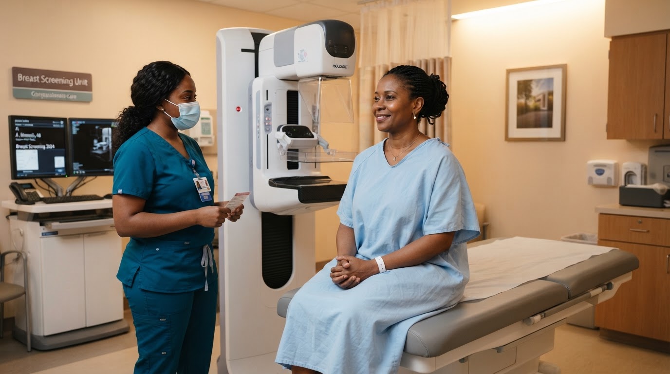
Mammography remains the undisputed gold standard for breast cancer screening. A mammogram is a specialized, low-dose X-ray of the breast that can detect microcalcifications and tiny tumors long before they can be felt during a physical exam. Modern digital mammography provides exceptionally clear images, allowing radiologists to spot the subtlest changes in breast tissue.
Guidelines regarding when to begin screening can vary, but most medical organizations recommend that women begin annual or biennial mammograms at age 40. Women with a strong family history of breast cancer or known genetic mutations (like BRCA1 or BRCA2) may need to begin screening even earlier. It is crucial to have a personalized discussion with your doctor to determine the screening schedule that is right for you.
While mammography is highly effective, it is not the only tool in our diagnostic arsenal. For younger women, or women who have dense breast tissue, mammograms can sometimes be harder to interpret because dense tissue and tumors both appear white on the X-ray. In these cases, a breast ultrasound is an invaluable complementary tool.
Breast ultrasound uses sound waves to create images of the internal structures of the breast. It is particularly useful for distinguishing between solid masses (which may be tumors) and fluid-filled cysts (which are almost always benign). When used in conjunction with a mammogram, ultrasound significantly increases the accuracy of the overall diagnosis.
Despite the proven benefits of screening, many women delay or avoid mammograms due to fear of pain, anxiety about the results, or simply a lack of time. As healthcare providers, we must acknowledge these concerns and strive to create a screening environment that is compassionate, supportive, and respectful of a woman's dignity.
At Crescent Radiology in Wuse, we have invested in modern imaging technology designed to provide accurate breast assessments with maximum patient comfort. Our female-led imaging teams understand the sensitivity of these procedures and are dedicated to guiding patients through the process with empathy and professionalism.
I urge every woman reading this to take control of her breast health. Perform regular breast self-exams to become familiar with your normal tissue, report any changes to your doctor immediately, and most importantly, adhere to your recommended screening schedule. Do not let fear prevent you from taking action; early detection is the greatest empowerment.
Related Articles

Taking Control of Cervical Health: Why Routine Pap Smears Are Non-Negotiable
Read Article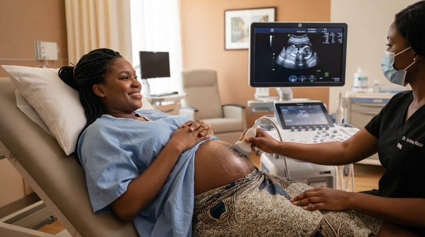
The First Glimpse of Life: The Indispensable Role of Obstetric Ultrasounds in Maternal Care
Read Article